Meng-qin Su ![]() ,
Wei Zhong,
Su-en Ren
,
Wei Zhong,
Su-en Ren
For correspondence:- Meng-qin Su Email: sumengqinss@hotmail.com Tel:+8637165662780
Received: 4 November 2015 Accepted: 11 May 2016 Published: 28 June 2016
Citation: Su M, Zhong W, Ren S. Dose-dependent protection of reseveratrol against spinal cord ischemic-reperfusion injury in rats. Trop J Pharm Res 2016; 15(6):1225-1233 doi: 10.4314/tjpr.v15i6.15
© 2016 The authors.
This is an Open Access article that uses a funding model which does not charge readers or their institutions for access and distributed under the terms of the Creative Commons Attribution License (http://creativecommons.org/licenses/by/4.0) and the Budapest Open Access Initiative (http://www.budapestopenaccessinitiative.org/read), which permit unrestricted use, distribution, and reproduction in any medium, provided the original work is properly credited..
Purpose: To examine the protective effects of resveratrol (RESV) against spinal cord ischemic reperfusion (SCIR) injury.
Methods: Forty-eight male rats were divided into six groups: sham-operated (control-I), SCIR-treated (SCIR-II), rats receiving 20 mg/kg of RESV with SCIR (RESV 20+SCIR-III), rats receiving 40 mg/kg of RESV with SCIR (RESV 40+SCIR-IV), rats receiving 60 mg/kg of RESV with SCIR (RESV 60+SCIR-V), and rats receiving 50 mg/kg of methylprednisolone (MP) with SCIR (MP + SCIR-VI), for 7 days prior to IR (pre-treatment) and 7 days after IR (post-treatment).
Results: The levels of oxidative markers (TBARS, MPO) and inflammatory markers (IL-1β, IL-6, TNF-α, and NF-p65) were concomitantly suppressed in RESV-treated rats, which showed improved locomotor function. A pronounced increase in the activities of antioxidant enzymes (SOD, CAT and GSH) was noted in the RESV group compared with the MP and SCIR groups. RESV and MP supplementation increased neuronal count with decreased nuclear degeneration. RESV (40 mg) exhibited greater protective effect than 20 mg and 60 mg of RESV and 50 mg of MP.
Conclusion: The results show the neurotherapeutic potential of RESV (40 mg) to attenuate oxidative stress and the inflammatory response to SCIR injury.
Introduction
Spinal cord ischemic reperfusion (SCIR) injury is a fatal postoperative complication of thoracic-abdominal aortic aneurysm surgery that contributes to paraplegia in about 10 % to 40 % of cases [1]. Paraplegia is a medical condition in which the sensory or motor part of the lower extremities is impaired due to spinal injury, resulting in long-term disability [2]. Numerous methods or techniques are recommended to treat paraplegic conditions, such as draining the cerebrospinal fluid, placement of shunts or performing a partial cardiopulmonary bypass, hypothermia, and administration of pharmacological products such as steroids and vasodilators [3]. However, these protective effects do not completely revert the paraplegic condition. Instead, these treatment regimens lead to various adverse events that may worsen the paraplegic condition.
Many researchers have focused on various natural products that may suppress spinal cord ischemic injury (SCII) and subsequently prevent the paraplegic condition. Several pathophysiological events, including excite-toxicity (glutamate-mediated), energy failure, inflammation, apoptosis, and oxidative stress, have been implicated in SCIR injury. Among these events, oxidative stress and inflammation are the major contributors and initiators of SCIR injury [4]. Therefore, choosing an antioxidant and an anti-inflammatory agent that alleviate SCIR injury would be a promising strategy. Recent studies have shown the protective effects of various natural antioxidant products against SCIR injury [5]. Methylprednisolone (MP) is a neuroprotective agent that possesses antioxidant and anti-inflammatory properties, and is recommended for treating spinal cord injuries [6]; thus, MP was used as a standard in the present study.
Resveratrol (RESV; 3,4',5-tri-hydroxy stilbene) is a phenolic compound commonly found in red wine/red grapes, soybeans and pomegranates [7]. RESV has two aromatic rings, with the free hydroxyl group contributing to its antioxidant and free radical scavenging activities [8]. RESV has been used as a potential drug for treating cardiovascular disease, diabetes, cancer, and various neurological disorders [9]. RESV exhibits neuroprotective activities due to its antioxidant, anti-inflammatory and anti-apoptotic properties [10].
RESV induces nitric oxide (NO) production, thereby acting as a vasodilatory agent [11]. Additionally, RESV readily crosses the blood-brain barrier, which favors its neuroprotective effects in various ischemic models. Several studies have shown the neuroprotective effects of RESV against SCII in various animal models using different treatment regimens [12]. However, to date, no studies have explored the long-term, dose-dependent protective effects of RESV against SCIR injury. Moreover, a treatment regimen using RESV (combination of both pre- and post-treatment) remains to be established. Therefore, in this study, we explored the effective dose of RESV against SCIR injury based on a combination of both pre- and post-treatment patterns (pre- and post-ischemic conditions) by assessing locomotor capacity, neuronal viability, and oxidative and inflammatory markers in a rat model.
Methods
Drugs and reagents
Resveratrol (RESV), Hematoxylin and eosin (H & E) and Methylprednisolone (MP) were procured from Sigma Aldrich (St. Louis, MO, USA). Paraformaldehyde, physiological saline (0.89 %), glycerol, sodium hydroxide, hydrogen peroxide were supplied by Lingjin Co. Ltd, Shanghai, China. Phosphate buffered saline (PBS) readymade buffer Xylene was bought from Beijing Zhongshan Golden Bridge Biotechnology Co. Ltd., Beijing, China.
Experimental animals
Healthy, adult male Sprague-Dawley (SD) rats weighing 300–320 g were used for the experiment. Rats were housed in a steel cage under a 12/12-h light/dark cycle. Food and water were given ad libitum. The Ethics Committee of China approved the animal experiments for the present study (protocol ref no. 02315), which were conducted according to the guidelines from the National Institutes of Health Guide for the Care and Use of Laboratory Animals.
SCIR injury was performed based on the method previously reported by Hwang [13] with slight modifications. Briefly, all rats were fasted overnight (12 h) and anesthetized with ketamine (50 mg/kg) injected intramuscularly. Body temperature was maintained at 37 °C using warming or heating pads and heating lamps. The hair around the neck region (carotid-inguinal) was shaved by placing the rats in the supine position. A polyethylene catheter (PE-50) was inserted into the tail artery to inject heparin and monitor the mean distal arterial pressure (MDAP). Thereafter, a Fogarty balloon catheter (Edwards Life Science, Shanghai, China) was inserted via the left femoral artery into the proximal descending thoracic aorta (approximately 11 cm from the insertion site), and the balloon was inflated with saline to induce spinal cord ischemia.
Proximal artery pressure was maintained at 80 mm Hg during aortic occlusion by draining the blood from the carotid artery into the external blood reservoir filled with heparinized saline. Immediate loss of pulse with decreased MDAP confirmed aortic occlusion. The balloon was deflated after 25 min of ischemia to initiate reperfusion by restoring blood flow. Then, the catheters were withdrawn and the rats were allowed to recover from anesthesia. All procedures were performed in sham-operated control rats, which served as negative controls, except for aortic occlusion. Mean arterial pressure, body temperature and heart beat (vitals) were continuously monitored before ischemic reperfusion.
Experimental protocol
Forty-eight male SD rats were divided into six treatment groups with eight rats per group. Rats received saline (sham-operated control-I), rats subjected to SCIR insult (SCIR-II), rats receiving 20 mg/kg b.wt of RESV dissolved in 2 % DMSO saline via i.p. (RESV 20+SCIR-III), rats receiving 40 mg/kg b. wt of RESV dissolved in 2 % DMSO saline via i.p. (RESV 40+SCIR-IV), rats receiving 60 mg/kg b. wt of RESV dissolved in 2 % DMSO saline via i.p. (RESV 60+SCIR-V) and rats receiving 50 mg/kg b. wt MP dissolved in saline via i.p. (MP + SCIR-VI) for 7 days prior IR (pre-treatment) and 7 days after IR (post-treatment).
Neurological assessment
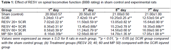
Locomotor function (hind limb movement) was tested by an independent observer blinded to the grouping and experimental design on days 1, 3, 5 and 7 after SCIR injury using the Basso Beattie Bresnahan (BBB) motor rating scale. The rating scale has 0 - 21 levels, where the value 0 (no detectable hind limb movement), 1 - 8 (rated for slight or higher movements of hindlimb joints), 9 (rated for dorsal stepping), 10 - 20 (rated for progressive improved walking ability) and 21 (normal movement).
After the last neurological assessment (on day 7), the rats in each group were subdivided into two subgroups for histological (n = 4) and biochemical analysis (n = 4). On the day 7, all rats were euthanized by i.p. pentobarbital sodium injection and spinal tissue were removed immediately and fixed in 4 % paraformaldehyde for morphological studies. Spinal tissue homogenate (10 %) was prepared in Tris-HCl buffer, centrifuged at 2500 g for 20 min and used for biochemical analysis.
Evaluation of edema (water content)
Edema (E) was detected using procedures previously published by Mdzinarishvili with a few modifications [14]. The wet and dry weights of spinal samples were measured to calculate the E (water content). E was calculated as the % difference between the two weights.
Lipid peroxidation and antioxidant enzyme assays
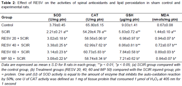
The levels of superoxide dismutase (SOD), catalase (CAT) and reduced glutathione (GSH), as well as the lipid peroxidation product (malondialdehyde, MDA), were measured in spinal tissue using commercial kits based on the manufacturer's protocols (Nanjing Jiancheng Bioengineering Institute, Nanjing, China). Bradford method was used to assess protein concentration.
Assay of myeloperoxidase activity
Myeloperoxidase (MPO) levels in spinal tissue were quantified by using MPO assay kit (Nanjing Jiancheng Bioengineering Institute, Nanjing, China). One unit of MPO activity was defined as the amount of enzyme degrading 1 mmol of peroxidase/min at 25 °C and expressed as Unit per gram (U/g) weight of wet tissue.
Assay of TNF-α, IL-1β, IL-6 and NF-κB p65 subunit
The concentration of various proinflammatory cytokines such as TNF-α, IL-1β, and IL-6 in spinal homogenate were assessed by commercially available ELISA kits (sandwich enzyme immunoassay) based on the manufacturer’s protocol (Thermo Fisher Scientific Inc, MA, USA). Similarly, the levels of the NF-κB free p65 subunit in the nuclear fraction (CellBiolabs Nuclear/Cytosolic Fractionation Kit) of the spinal tissue was determined by ActivELISA (Imgenex, San Diego, USA) kit.
Histopathological examination
Four rats in each group were euthanized and the spinal cord (tissue) were removed, fixed in 10 % buffered formalin for 24 h and embedded in paraffin. Transverse sections were cut at a thickness of 4 µm and stained with H & E (100x) .Images were captured with a high-resolution digital camera attached to a Nikon Eclipse 80i microscope (Nikon Co., Tokyo, Japan) and analyzed by Image Pro Plus software.
Statistical analysis
All values are explicated: mean ± standard deviation (SD). Variation between each experimental groups were measured by one way analysis of variance (ANOVA) using the software SPSS 21 version (SPSS Inc., Chicago, USA) and least significant difference (LSD) was determined using post-hoc multi-comparison test. P < 0.05 was deemed statistically significant.
Results
Effect of RESV on locomotive function
Movement (motor function) of hind limbs were normal in the sham-operated control rats (). However, the SCIR treated animals showed a significant reduction in hind limb movement, which depicts the loss of several neurons. After supplementation with RESV and MP, hind limb movement was substantially improved, thus lowering the incidence of neurological deficits.
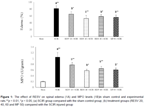
Effect of RESV on spinal edema
As shown in A, spinal E values were dramatically higher (81.35 %) in the SCIR group compared with the sham-operated control group. RESV (20, 40 and 60 mg/kg) groups showed lower spinal E values (65.12, 52.56 and 58.34 %, respectively) than SCII group; MP treatment resulted in an E value of 55.82 %, similar to that of RESV 40 group.
Effect of antioxidant and lipid peroxidation products (MDA)
The levels of antioxidant and lipid peroxidative products (MDA) in SCIR-injured rats were examined to determine whether RESV administration protected spinal tissue against oxidative stress (). The spinal SOD, CAT, and GSH activities were is substantially lower (p < 0.05) in the SCIR group than in the sham-operated control group, whereas the MDA levels were markedly elevated (p < 0.05). Treatment with RESV significantly improved (p < 0.05) SOD, CAT and GSH activities as well as concomitantly attenuated the MDA level in spinal tissue. However, the MP group showed a slight improvement in antioxidant activity and partially reverted the MDA levels. RESV 40 (p < 0.01) showed higher antioxidant activity than other RESV doses or MP groups.
Effect of RESV on neutrophil infiltration (MPO)
To evaluate the effect of RESV on neutrophil activation and infiltration, MPO activity was determined (B). Compared with the SCIR group, the sham-operated group showed a decline (p < 0.05) in MPO activity. Supplementation with RESV and MP markedly attenuated the MPO activity in a dose-dependent fashion.
Effect of RESV on activities of inflammatory markers
The levels of these inflammatory markers in the spinal tissue of SCIR induced rats were higher (p < 0.01) as compared with sham control rats. Equivalence with SCIR induced rats, RESV and MP treatment (p < 0.01) lowered the concentrations of these pro-inflammatory cytokines, thus suggesting its anti-inflammatory potential.
Effect of RESV on spinal section with hematoxylin and eosin staining
Control showed normal architecture of neuron with a prominent nucleus (Plate 2A). The spinal section of SCIR treated rat portrait a higher degenerated neurons (hemorrhage and congestion) with widespread edema, increased cytoplasmic vacuolization and disintegrated nucleus (2B). However, RESV-administered rats (2C - E) showed fewer degenerated neurons (hemorrhage and congestion) compared with SCIR-injured animals, confirming the neuroprotective effects of RESV. Similarly, MP treatment (2F) also showed a significant restoration in neuronal structure, but still mild edema accompanied with few degenerated neurons.
Discussion
Paraplegia is a fatal complication resulting from SCII during thoracic-abdominal aortic aneurysm surgery [1]. Several pathophysiological events, including excitotoxicity (glutamate-mediated), energy failure, inflammation, apoptosis, and oxidative stress, have been implicated in SCIR injury [4,5]. Among these, oxidative stress and inflammation are the major contributors and initiators of SCIR injury [4-6]. Therefore, in the present study, we examined the protective effects of RESV against SCIR injury in a dose-dependent manner by evaluating various oxidative stress and inflammatory markers using MP as a standard.
Assessment of locomotor behavior (hind limb movement) is an important measure of long-term functional recovery after SCIR injury and is a pivotal tool for determining the neuroprotective efficacy of various drugs. Our results clearly show the neuroprotective efficacy of both RESV and MP based on markedly increased BBB scores. This result reflects improved hind limb movement by preventing neuronal loss and mitigating the incidence of neurological deficits. Liu et al also suggested that RESV can significantly lower neurological deficits in a SCIR-injured rabbit model [15].
Morphological abnormalities of spinal tissue are indirectly assessed by evaluating E based on dry and wet weights. During SCIR induction, a substantial reduction in blood flow was observed (due to aortic occlusion), followed by reperfusion (increased blood flow), which alters the electrolytic balance (pumps) and various metabolic processes in spinal tissue, leading to elevated water movement and eventually resulting in E [13,15]. Due to the vasoprotective effects of RESV [16], the electrolytic balance was significantly maintained, thus attenuating E. Our results are in agreement with those of Yang et al, who showed that RESV treatment can revert E successfully in the SCIR-injured rat model [17]. Similarly, Ren et al also inferred that RESV can effectively reduce E under ischemic conditions [18].
Considerable evidence indicates that overproduction of free radicals (oxidative stress) and the subsequent inflammatory response under ischemic conditions are major factors contributing to various pathophysiological changes during SCIR injury [13,19]. Neural tissues are highly susceptible to free-radical damage due to an increased metabolic rate and lipid content. During SCIR, many free radicals are generated; to counter the excess free radicals the endogenous antioxidant defense system, which includes SOD, CAT and reduced GSH, is upregulated by various factors to trigger the Nrf2-dependent pathway. However, high levels of free radicals suppress normal antioxidant levels and ultimately contribute to oxidative stress. Therefore, SOD, CAT and GSH levels were significantly lower in SCIR-injured rats.
Moreover, levels of the lipid peroxidation product MDA were elevated in the SCIR group, contributing to oxidative stress. RESV-supplemented animals exhibited improved SOD and CAT activities and higher GSH levels, as well as substantially reduced MDA levels, in spinal tissue. Overall, RESV 40 showed better antioxidant activity compared with other RESV dosages or the MP group. Due to the free hydroxyl group, RESV may act as an antioxidant and trigger endogenous antioxidant levels to revert oxidative stress conditions by maintaining a balance between antioxidants and free radicals and activating the Nrf2/HO-1 pathway [20]. Furthermore, RESV has been reported to be a xanthine oxidase inhibitor and halt the synthesis of hypoxanthine and free radicals (ROS) under ischemic conditions [21].
Inflammation is the crucial event that is responsible for the neurological damage (SCI) due to ischemic reperfusion and thus has been proven to be a promising target for a therapeutic approach after SCIR injury [21]. Neutrophil and neuroglial activation, mobilization and infiltration are well-documented key factors during SCIR that initiate the inflammatory cascade by triggering NF-κB and subsequently increasing various pro-inflammatory cytokines such as tumor necrosis factor-α, IL-1β and IL-6 [22]. Hence, the inflammatory status of spinal tissue was indirectly evaluated by measuring MPO activity. Several reports have indicated a strong correlation between MPO levels and the severity of ischemic reperfusion injury [23]. Lafci et al also showed that MPO activity was significantly increased after SCIR injury in a rat model [24]. Similarly, in the present study, MPO activity in spinal cord tissue was significantly increased in response to ischemic reperfusion. The increased MPO activity was markedly attenuated by the RESV and MP treatments, indicating the antioxidant and anti-inflammatory activities of RESV. Renaud and Martinoli argued that treatment with RESV substantially reduces microglial activation and significantly inhibits the infiltration of neutrophils, thus diminishing MPO levels under ischemic conditions [25].
Overexpression of NF-κB has been positively correlated with a higher degree of ischemia-induced neuronal injury [15]. Levels of pro-inflammatory cytokines such as TNF-α, IL-1β, IL-6, and nuclear factor NF-p65 were dramatically increased in the SCIR rats due to excessive production of free radicals which, in turn, triggered the neuroinflammatory cascade by activating various cytokines. Compared with the SCIR-injured rats, the RESV and MP treatments substantially suppressed the concentrations of these pro-inflammatory cytokines, indicating their anti-inflammatory potential. Our results are supported by those of Smith et al., who evaluated inflammatory chemokine concentrations (interleukin-1β, IL-6 and TNF-α) and reported elevated levels of these inflammatory cytokines after SCIR [26]. Furthermore, RESV can effectively improve cerebral blood flow by crossing the blood-brain barrier and exhibits protective effects against ischemic injury by decreasing glial cell activation and thus positively alleviating oxidative stress and the inflammatory cascade.
RESV can also influence SIRT1 expression indirectly at the transcriptional level, block the translocation of NF-κB (p65) from the cytosol to the nucleus, and inhibit the production of various pro-inflammatory cytokines, especially IL-1β, IL-6 and TNF-α. Moreover, RESV augments endogenous antioxidant activities, thus inhibiting activation of the inflammatory cascade [15].
Sham-operated control rats showed normal neuronal architecture with prominent nuclei. However, SCIR-injured rats had higher numbers of degenerated neurons (hemorrhage and congestion) with widespread E, increased cytoplasmic vacuolization and disintegrated nuclei due to increased oxidative stress and cell damage by lipid peroxidation (MDA). As explained previously, RESV and MP concomitantly improved the antioxidant status and attenuated MDA levels substantially, indicated by the fact that RESV- and MP-administered rats displayed significantly fewer degenerated neurons (hemorrhage and congestion) than SCIR-injured animals, validating the neuroprotective effects of RESV.
Liu et al [15] also demonstrated that RESV treatment normalized the neuronal count and function due to antioxidant, anti-inflammatory and anti-apoptotic activities. RESV (40 mg) exhibited the highest neuroprotection compared with MP due to its free radical-quenching activity. Our results are in agreement with the findings of Ates et al and Yang et al [12,17]. The major limitation of the current study is that bioenergetics parameters and apoptosis were not evaluated to confirm secondary energy failure and its association with RESV treatment. Additionally, the reason for the improved activity of RESV (40 mg) should be explored using different molecular techniques.
Conclusion
Both RESV and MP exhibit neurotherapeutic properties, reflected in various aspects, including substantially reduced E, oxidative stress and inflammation. Furthermore, RESV and MP exhibit spinal protective properties by concomitantly reverting histopathological abnormalities and improving the number of neurons. Thus, it is proposed here that RESV (40 mg) can be used as a neurotherapeutic agent to treat SCIR injury and lower the risk of paraplegia significantly.
Declarations
Acknowledgement
References
Archives


News Updates